Gram Stain Lab
Background Info
Wikipedia > Gram-negative bacteria
Wikipedia > Gram-positive bacteria
How Gram Staining Works > Simplified
To understand gram staining you must be familiar with the structure of the bacteria they are used on.
Gram positive bacteria have a single thick cell wall made of peptidoglycan (50-90% of cell wall). This thick wall stains purple and remains purple throughout the gram staining test.
Gram negative bacteria have a thinner layer of peptidoglycan (10% of cell wall), and an additional outer membrane which contains lipids. The outer membrane of gram negative bacteria will stain purple also but it falls off during the gram stain test. Its 2nd wall is what gets stained pink. We stain it pink so we have a color contrast to look at.
Virtual Lab
Gram Staining Pitfalls
It is clear that the decolorization step is the one most likely to cause problems in the gram stain. But other problems can arise too (some listed below). Think about the purpose of each step and answer the questions on your virtual lab capture sheet.
Slide over-decolorized: Gram positive (purple) bacteria will appear gram negative (pink).
Slide under-decolorized: Gram negative (pink) bacteria will appear gram positive (purple).
Excessive heat during fixation: Heat fixing the cells, when done to excess, alters the cell morphology and makes the cells more easily decolorized.
Excessive washing between steps: The crystal violet stain is susceptible to wash-out with water (but not the crystal violet-iodine complex). Do not use more than a 5 second water rinse at any stage of the procedure.
Excessive counterstaining: As the counterstain is also a basic dye, it is possible to replace the crystal violet—iodine complex in gram- positive cells with an over-exposure to the counterstain. The counterstain should not be left on the slide for more than 60 seconds.
Bacteria Analysis
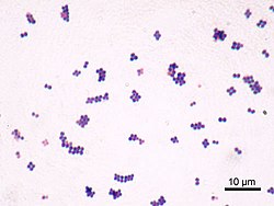
1. Describe the image using shape, arrangement, and gram stain of the bacteria.

2. Describe the image using shape, arrangement, and gram stain of the bacteria.

3. Describe the image using shape and gram stain of the bacteria.

4. Describe the image using shape and gram stain of the bacteria.

5. Below is a Gram Stain test of unknown bacteria. Would this test result be considered valid? Explain why or why not.


Positive Control Negative Control
Unknown/ Test Sample
6. a) If the results below were obtained after performing a gram stain, what could be concluded about the staining process?
b) What is most likely the reason we see this problem? (Hint: Look at the Gram Staining Pitfalls section)
c) Which step of the gram staining process is most likely affecting the test results?


Positive Control Negative Control

Unknown/ Test Sample